How CT scans work
Professor John Slavotinek, Dean of Clinical Radiology at the Royal Australia and New Zealand College of Radiologists explains.
Health Agenda magazine
July 2016
The scientists behind this 1972 invention, Godfrey Hounsfield and Allan Cormack, were awarded the Nobel Peace Prize in 1979 for their contributions to medicine and science.
Here, we explain the why CT scans are increasingly used as a diagnostic tool.

What is it?
CT stands for computed tomography, which uses an X-ray beam to record cross-sectional images of the inside of the body including bones, soft tissue and blood vessels. This provides detailed information for a range of conditions and can also detect more subtle abnormalities than traditional X-rays. CT scans are also able to confirm if a previously treated disease has recurred.
What does it do?
CT scans are very commonly used in trauma, for assessing injuries resulting from a car accident, for example. Other purposes include diagnosis and follow-up of tumours, and minimally invasive therapies. CT is an effective means of guiding biopsy procedures performed by radiologists.
Why would I have it?
If you’ve suffered a traumatic injury, CT scans will provide detailed information about the extent of the damage. Radiotherapy patients can receive beams to shrink their tumours without the need for external markers, and even steroid injections for pain relief can be precisely guided using CT.
The process
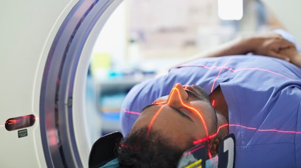
Patients lie still on a flat bed for a CT, but this doughnut-shaped machine is not as confining as an MRI and is quieter. Often a contrast agent is used, especially for trauma scans. This is injected with a fine needle and causes little discomfort. Common side effects may include a temporary warm sensation over the entire body or a temporary unpleasant taste in the mouth. A straightforward CT scan can be conducted in as little as 10 seconds.