How MRIs work
Professor John Slavotinek, Dean of Clinical Radiology at the Royal Australia and New Zealand College of Radiologists explains.
Health Agenda magazine
July 2016
Magnetic resonance imaging (MRI) is a powerful diagnostic tool that uses a magnetic field and radio waves to produce highly detailed images of the inside of the body.

What is it?
MRI was invented by Armenian-American doctor Raymond Damadian in the 1970s, and can be used to assess the condition of organs, the spine, bones and soft tissue including ligaments and cartilage.
Unlike standard X-rays, MRI doesn’t use ionising radiation, which is known to be harmful with prolonged exposure. Even so, it isn’t suitable for everyone – for instance people who’ve been fitted with certain types of pacemaker.
What does it do?
MRI is used to diagnose conditions, determine their extent and guide subsequent treatment. It allows doctors to identify the stage of a disease and plan medical intervention. In a cardiac MRI, for example, doctors can view the movement of a beating heart to assess cardiovascular disorders.
Why would I have it?
You may be referred for an MRI scan for any condition involving the brain, such as stroke or tumour, as well as other neurological conditions. If you have symptoms relating to your organs, spine, or musculoskeletal structures such as joints, an MRI provides an accurate picture of what’s going on.
The process
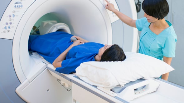
After being screened for implants that may be affected by the MRI’s magnetic fields, you’ll be asked to lie on a flat bed, which then slides into a tunnel-like machine.
Depending on what is being scanned, your entire body may not be inside the machine, but to get the best result you need to keep as still as possible. As the machine is quite noisy you may be given headphones. A typical MRI takes 15-20 minutes.